

This story is a sad example about how illiterate bureaucracy, for quite poorly specified reason, is capable to stop a pioneering research and to delay by many years the opportunity for progress of important area of science.
1. First steps of technology development
This method was one of the first digital microscopy methods, which main goal was the registration of the stable and reproducible profile of the cortical area of human brain. At the time of its development, computers were very simple and not at all powerful, and even such terms as “medical informatics” and “digital pathology” did not exist. However, considering modern definition of digital pathology as an image-based information environment enabled by computer technology, our set of programs and algorithms was truly pioneering technology. Due to it’s stability and reproducibility, the procedure was given a name “Automatic Morpho-Corticography”, of MCG.
I should start the review of MCG technology from a short tribute to my Alma Mater – the N. I. Pirogov’s Second Moscow Medical School (presently – Pirogov’s Russian National Research Medical University). The reason for that is not just a mere politeness or pride. The former Rector (or Dean, in American English) of the 2nd Moscow Medical School, prof. Yu. M. Lopuhin, who is better known for his contributions to transplantology and hemosorbtion, had an amazing ability to foresee the future of Medicine. He and prof. S.A. Gasparian were among the first people in Russian (or rather “Soviet”) medical science who understood the significance of computers and software engineering in medicine (“medical cybernetics”, as they used to say at that time). Many years before it became well known and widely used, they were able to understand the importance of medical imaging, computerized microscopy and computer-assisted diagnosis. Thus, in 1973, they established in the Second Moscow Medical School the first in the world Department (or “Faculty”) of Medical Cybernetics. Medical students, in addition to medical education, were able to get training in programming, computer science, image processing and other fields related to the application of computers in medicine.
Importantly, the teaching process was organized in such a way, that starting from the second year of education students were able to participate in solving practical scientific problems using the most powerful computers and computerized equipment available at that time. This opportunity was open even for students enrolled in “general” medical program. So, I wrote my first code for automatic scanning and segmentation of histological specimens in the Laboratory of Prof. V.E. Nemirovsky, in 1974. I was a third-year student and started working with first two computers I ever saw in my life: Wang 720C and PDP-12. Computers were just unpacked and installed, and radiated this unique head-spinning smell of brand-new “western” electronics, which very few could experience first hand at that time. I said “code”, since high-level language for Wang-720C was not available: the program had to be developed in hexadecimal “auto-code” (0407 – “GoTo”, 0408 – “Label”, 0300 – “+” etc.). Given this experience, I never take C++, Python and Matlab for granted.
After graduation I was fortunate to start working in the All Union Research Center of Mental Health, which had two relatively independent institutions: Institute of Clinical Psychiatry and Moscow Institute of Brain Research. Exceptionally well funded, the Center had unique equipment, including several computers from USA, many German light microscopes, two unique image analysis systems TAS from Ernst Leitz Wetzlar, and several electron microscopes by Fillips. It is interesting to note, that Moscow Brain Institute was practically the only place in the Soviet Union, where cytoarchitectonic studies of human brain were traditionally conducted. Traditions of such studies are due to German scientist Oscar Vogt, who founded this Institute in 1924.
Initially, the decision to organize the Laboratory for Study of Lenin’s brain happened shortly after the his death, and that was the place where Lenin’s brain was carefully stored and meticulously studied. The goal of the Laboratory was to explore Lenin’s brain and to discover the reason of his geniality. Invited by Sovnarkom (Soviet of Peoples’s Commissars), Oscar Vogt personally brought from Germany a lot of equipment, mainly – for histological processing of the brain, including serial sectioning, and microscopy. In 1928, the neuroanatomical laboratory of Vogt was reorganized into the Moscow Brain Research Institute, and Vogt became its first director. Structured collecting and mapping of the brain started. Later many more brains of famous and prominent people were collected in the Institute, and it became the center of studies of human brain architecture with explicit goal to understand the structural basis of human intelligence. It is not surprising that Institute brain collection included the brains of prominent Russian neuroscientists, such as neurologist, G.I. Rossolimo, physiologist, I.P. Pavlov‚ neurologist M. B. Kroll , psychiatrist P. B. Gannushkin psychologist L.S. Vygotsky. There were also brains of poet V.Mayakovsky, physicist L.D. Landau, scientist E.K.Tsiolkovsky, writer A.M.Gorky.
Of course, the goal of the Moscow Brain Institute was not achieved. It is still a big challenge to show how cortical structure and spatial organization of neurons can be connected to higher cognitive brain function. However, many important results were generated, including development by S.A. Sarkisov and colleagues of modified cytoarchitectonic Brodmann’s map [S.A.Sarkisov et al., “Citoarchitecture of the cortex of human brain”, Moscow, 1949] .
2. Design of the MCG technology and implementation conditions
For its time, it was truly pioneering technology. It was based on algorithms of Mathematical Morphology (J. Serra, CMM, France), implemented in the texture analysis sytsem TAS, by Ernst Leitz, Wetzlar (see fig. 1)

Fig. 1: Brief technical description of automatic microscopy system Leitz TAS (1980-1982).
Once again, our method was one of the first digital microscopy method, developed for registration of the stable and reproducible profile of the cortical area of human brain. We defined “stability” as obtaining statistically indistinguishable result when the measurement was repeated in the same area, which was achieved by adaptive segmentation algorithm based on measuring of mean gradients distribution, and rather sophisticated methods of elimination of artifacts, vessels and glia. Reproducibility was defined as obtaining the same profile pattern and average values measured in the same area, but different set of serial sections, what was achieved by satisfying ergodicity requirements (see, for example “Ch. Lantuejoule, Ergodicity and integral range, J. of Microscopy, 161, 3, 1991. Practically it meant processing very significant volume of tissue in each area, at least within 2 mm of section space at ~<0.5 mkm resolution). Starting from step 4 (below) it was fully automatic.
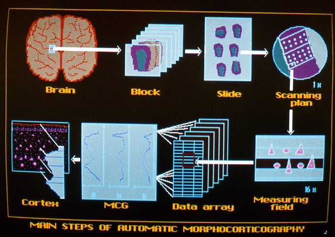
Fig. 2: MCG processing pipeline.
The method was called “Morphocorticography” because in a substantial number of cases it revealed a pattern of local minima and maxima, specific for a given individual. This pattern reproduced itself in different areas of the same individual, but not in another individual.
All details of the method, including MM notations of image processing steps, were published between 1982 and 1992 in Russian (translated in English, Istomin VV, Neuropatol. and Behav. Physiol.,1986, UDC 616.831.31-076.5), English (V. Istomin, K. Amunts, Vision & Voice Magazine, 6, 2, 1992) and German (Istomin V., Amunts K., Build und Ton, 43, 1, 1990 and Amunts K, Istomin V., Vision & Voice Magazine, 6, 1, 1992). Unfortunately, these journals were not very well known, and probably do not even exist anymore.
Thank you for paying tribute and preserving memory of yhe people who evidently have contributed so much for the scientific development!
At the place and time where every substantial and good deed must have been done contrary to the prevailing standard, those scientists and truly gifted administratirs had the strength and dedication to carry out their vision! The following generations of the medical practitioners and researchers will carry on!